"Advanced imaging technology allows us to see what traditional X-rays cannot. At Cookeville Oral Surgery, our 3D cone beam CT scanner gives us unmatched visualization for precise diagnosis and treatment planning."
What Is 3D Cone Beam CT Imaging?
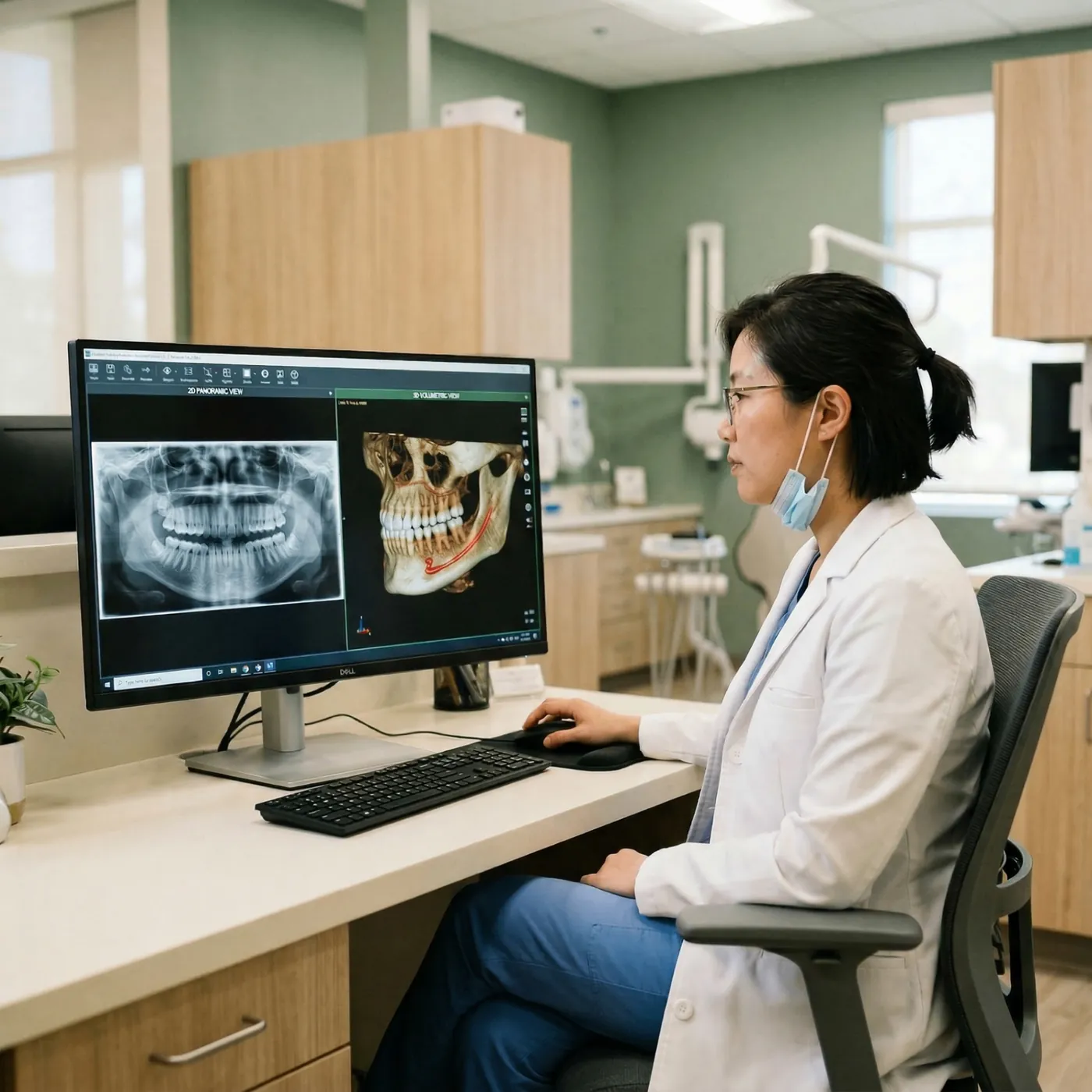
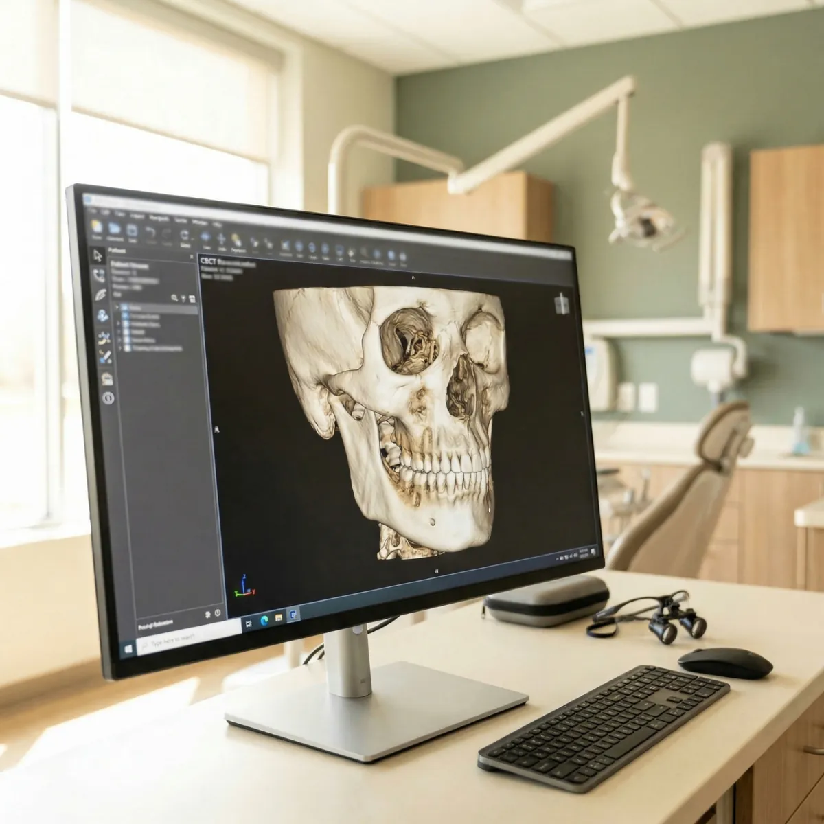
Cone beam computed tomography (CBCT) is a specialized type of X-ray technology that produces detailed 3D images of your teeth, jawbone, nerve pathways, and soft tissues in a single scan. Unlike traditional 2D dental X-rays, CBCT provides a complete three-dimensional view of your oral and maxillofacial structures.
Our state-of-the-art CBCT system captures crystal-clear digital images while minimizing your radiation exposure compared to traditional hospital CT scanners. The scan is quick, painless, and completed right here in our Cookeville office — no hospital visit required.
This technology enables us to perform a wider range of diagnoses and develop more precise treatment plans, often reducing the need for multiple visits and improving treatment outcomes.
Benefits of 3D Imaging
Precise 3D visualization of teeth, bone, and nerves
Lower radiation than traditional CT scans
Fast scan completed in under 30 seconds
Improved accuracy for implant placement
Better surgical planning and outcomes
No hospital visit required
Customizable field of view for targeted scanning
Easy image sharing with referring doctors
Our CBCT technology provides the detailed anatomical information our surgeons need to plan your treatment with confidence and precision.
How We Use 3D Imaging
Dental Implant Planning
3D imaging allows us to assess bone density, volume, and the precise location of nerves and sinuses before placing implants. This ensures optimal implant positioning for long-lasting results.
Jaw Surgery Planning
For corrective jaw surgery, CBCT provides detailed views of jaw alignment, airway dimensions, and facial structure to plan the most effective surgical approach.
Wisdom Teeth Evaluation
We can visualize the exact position of impacted wisdom teeth relative to nerves and sinuses, allowing for safer and more predictable extractions.
Pathology & Tumor Assessment
3D imaging helps identify and evaluate cysts, tumors, infections, and other pathological conditions within the jawbone and surrounding structures.
What to Expect During Your Scan
No special preparation is needed for a CBCT scan. You may be asked to remove jewelry, eyeglasses, or any metal objects near your head and neck area. Our team will position you comfortably in the scanner.
3D Imaging vs. Traditional X-Rays
Traditional dental X-rays provide a flat, two-dimensional view of your teeth and jaw. While useful for basic diagnostics, they can miss important details by overlapping structures.
Our 3D CBCT scanner captures a complete volumetric image, allowing our surgeons to view your anatomy from any angle and at any depth. This means more accurate measurements, better identification of conditions, and more predictable treatment outcomes.
Despite providing far more diagnostic information, our CBCT system uses significantly less radiation than a traditional hospital CT scan, and the focused beam technology allows us to scan only the area of concern.